Diencephalon
Model
Anatomical regions
Diencephalon
Diencephalon
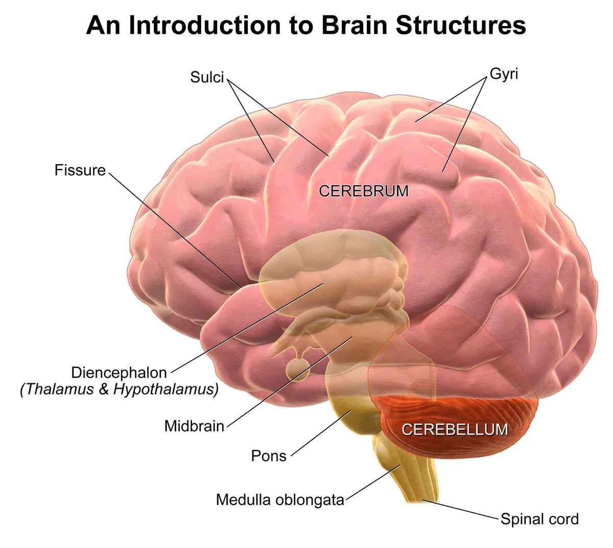
Lies between the cerebral hemispheres around the third ventricle; it comprises the thalamus, hypothalamus, epithalamus and subthalamus.
Functions
- Thalamus: relay of all sensory pathways except olfaction to the cortex
- Hypothalamus: regulation of body temperature, feeding, circadian rhythms and the hypothalamic–pituitary axis
Description
Lies between the cerebral hemispheres around the third ventricle; it comprises the thalamus, hypothalamus, epithalamus and subthalamus.
Key structures
Thalamus
- Name
- Thalamus
- Role, notes
- Relay of sensory and motor input to cortex (ventral posterior, lateral and medial geniculate nuclei)
Hypothalamus
- Name
- Hypothalamus
- Role, notes
- Endocrine control through the pituitary, temperature, feeding, circadian rhythm
Epithalamus (glandula pinealis)
- Name
- Epithalamus (pineal gland)
- Role, notes
- Melatonin secretion
Subthalamus
- Name
- Subthalamus
- Role, notes
- Subthalamic nucleus: part of the basal ganglia loop; lesions cause hemiballism
Corpora mammillaria
- Name
- Mammillary bodies
- Role, notes
- Part of the Papez circuit; affected in Wernicke–Korsakoff syndrome
| Terminologia Anatomica | Name | Role, notes |
|---|---|---|
| Thalamus | Thalamus | Relay of sensory and motor input to cortex (ventral posterior, lateral and medial geniculate nuclei) |
| Hypothalamus | Hypothalamus | Endocrine control through the pituitary, temperature, feeding, circadian rhythm |
| Epithalamus (glandula pinealis) | Epithalamus (pineal gland) | Melatonin secretion |
| Subthalamus | Subthalamus | Subthalamic nucleus: part of the basal ganglia loop; lesions cause hemiballism |
| Corpora mammillaria | Mammillary bodies | Part of the Papez circuit; affected in Wernicke–Korsakoff syndrome |
Boundaries and structure
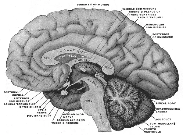
The diencephalon lies between the hemispheres around the third ventricle and comprises the thalamus, hypothalamus, epithalamus (pineal gland) and subthalamus. The pituitary hangs below the hypothalamus on the pituitary stalk, in the sella turcica of the sphenoid, and has a dual embryological origin (anterior lobe from Rathke’s pouch, posterior lobe from the diencephalon).[1]
Connections and function
The thalamus relays and modulates most information ascending to the cortex and takes part in thalamocortical loops governing consciousness and sleep. The hypothalamus, with thousands of distinct cell types, integrates peripheral, environmental and neural signals to regulate temperature, feeding, sleep and innate behaviour, and controls the pituitary.[2][3][4]
Thalamus: cell types, firing modes, drivers and modulators
At 4 weeks the neural tube forms three vesicles; by week 5 the prosencephalon has split into telencephalon and diencephalon, which differentiates into thalamus and hypothalamus. Thalamic neurons are born around the third ventricle and migrate laterally along radial glia, the earliest cells ending up most lateral; separate nuclei become discernible at gestational weeks 8–16, and thalamic development precedes that of the telencephalon.[2]
The thalamus has two basic cell types. Projection neurons are excitatory and glutamatergic, sending axons to the cortex, striatum and amygdala and giving collaterals to the reticular nucleus as they leave. Inhibitory GABAergic interneurons stay within their nucleus. Unlike the cortex, the thalamus has no recurrent intrathalamic excitation, which may restrict it to organising how a signal is perceived rather than modifying the signal itself.[2]
Projection neurons fire in burst or tonic mode. In burst mode, under strong hyperpolarisation, slight depolarisation opens T-type Ca²⁺ channels and triggers a burst of action potentials; slow inactivation (100 ms or more) limits bursts to a few per second, and the output does not faithfully reflect the input, although it is a sensitive detector of change. In tonic mode, from a depolarised state, firing frequency is proportional to input, so information passes with fidelity. During sleep thalamic neurons are mostly in burst mode; attending to facial sensation, for example, may switch the ventral posteromedial nucleus to tonic mode.[2]
In specific nuclei, afferents are drivers or modulators. Drivers carry the content via glutamatergic synapses: retina to lateral geniculate, gracile and cuneate nuclei to the ventral posterolateral nucleus. Modulators come from cortical layer VI, the basal ganglia, cerebellum, brainstem reticular formation and inhibitory intrathalamic neurons, using glutamate, GABA, serotonin, dopamine, acetylcholine and noradrenaline. Modulators far outnumber drivers and decide whether, and in what form, the content reaches the cortex.[2]
Thalamic nuclei and lesions
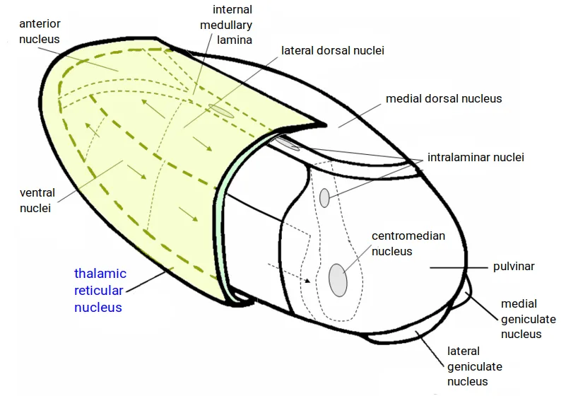
The thalamus has about 30 nuclei, not easily seen on routine MRI and named differently across atlases. The anterior nucleus (three subnuclei) belongs to the Papez circuit: hippocampus, fornix, mammillary bodies, mammillothalamic tract, anterior nucleus, cingulate cortex, entorhinal cortex and back to the hippocampus; it also organises sleep spindles and memory consolidation. In the ventral tier the nuclei run front to back: ventral anterior and ventral lateral receive cerebellar input and serve the motor system; the ventral intermediate nucleus relates to deep sensation and tremor; the ventral posteromedial (face) and posterolateral (body) nuclei receive sensory tracts and project to the sensory cortex. The lateral and medial geniculate nuclei receive visual and auditory input.[2]
The pulvinar is the largest nucleus and the one that has grown most in evolution; an association nucleus linked to posterior parietal, occipital, insular, cingulate and anterior temporal cortex, it serves multimodal processing and visual attention. The reticular nucleus covers the lateral thalamus like a blanket and is the only nucleus without excitatory output: it projects back onto the thalamus with inhibitory synapses, serving selective attention in wakefulness and sleep spindles in sleep. The dorsomedial nucleus, largest on the medial surface, projects to prefrontal, cingulate and insular cortex and receives the amygdala; it is involved in affect, memory and consciousness, and bilateral lesions can cause coma. The centromedian nucleus, an intralaminar nucleus, projects mainly to the striatum and is part of the reticular activating system.[2]
Korsakoff syndrome, from vitamin B1 deficiency in alcohol use disorder, causes anterograde and retrograde amnesia with the most marked degeneration in the anterior nucleus. Fatal familial insomnia, a rare autosomal dominant prion disease, preferentially destroys the dorsomedial and anterior nuclei, and loss of slow-wave sleep is among its earliest signs. Thalamic vascular lesions cause location-dependent memory disturbance, reduced consciousness or coma, aphasia, hemiataxia, pain syndromes and sensory loss. Acquired thalamic lesions do not usually cause epilepsy; the exception is neonatal thalamic injury, linked to epileptic encephalopathy with spike-wave activation in sleep (DEE-SWAS).[2]
Functions
- 01Thalamus: relay of all sensory pathways except olfaction to the cortex
- 02Hypothalamus: regulation of body temperature, feeding, circadian rhythms and the hypothalamic–pituitary axis
Regions
References
Sources for the values and descriptions on this page, indexed in PubMed (US National Library of Medicine).
- [1]Anatomy of the pituitary glandSav A et al., Best practice & research. Clinical endocrinology & metabolism 2025PMID 40500594
- [2]The thalamus: Structure, function, and neurotherapeuticsMarcuse LV et al., Neurotherapeutics 2025PMID 39956708
- [3]Thalamocortical interactionsSherman SM, Current opinion in neurobiology 2012PMID 22498715
- [4]The structural and functional complexity of the integrative hypothalamusFong H et al., Science 2023PMID 37883552
- [5]Automatic parcellation of human cortical gyri and sulci using standard anatomical nomenclatureDestrieux C et al., 2010PMID 20547229Supports: Nomenclature of cortical gyri and sulci
- [6]Neuroanatomy, Cerebral HemisphereStatPearls (NCBI Bookshelf)PMID 31747196
- [7]Neuroanatomy, CerebellumStatPearls (NCBI Bookshelf)PMID 30844194
- [8]Current Controversies on Wernicke's Area and its Role in LanguagePMID 28656532Supports: Wernicke’s area
- [9]The human brain in numbers: a linearly scaled-up primate brainHerculano-Houzel S, 2009PMID 19915731