細胞與細胞器
模型
懸停或輕觸可檢視名稱,點選可閱讀說明;點選圖例中的名稱可飛至該結構。按組織學比例以演算法生成,顏色遵循插圖慣例。
描述
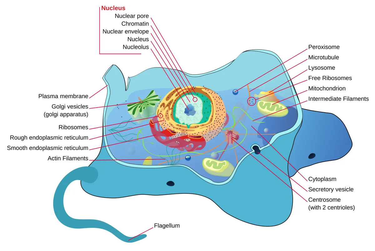
真核細胞由包圍細胞質和細胞核的細胞膜構成。細胞質中有膜性細胞器(內質網、高基氏複合體、粒線體、溶體、過氧化體)、非膜性細胞器(核糖體、中心體)和細胞骨架。每種細胞器都是專門的區室,協同完成合成、運輸、產能、降解和分裂。
細胞膜
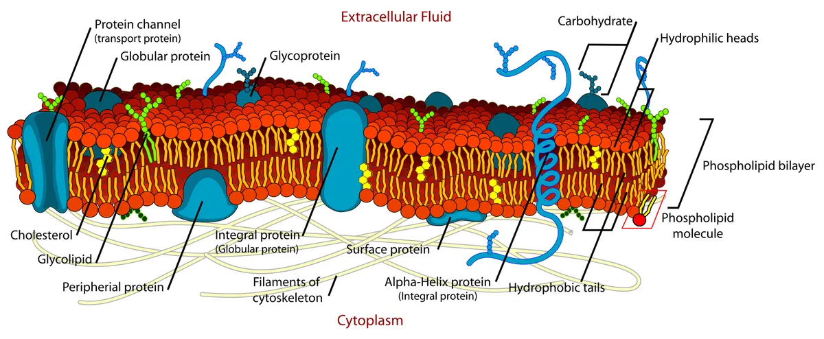
細胞膜是厚約 7–8 nm 的磷脂雙分子層,疏水尾朝內,其間鑲嵌膽固醇和蛋白質。按照流動鑲嵌模型,脂質和許多蛋白質可在膜平面內側向擴散;跨膜蛋白充當通道、泵、載體和受體,外周蛋白附著在膜的兩側。兩層並不相同:磷脂醯絲胺酸位於內層,它一旦暴露到外側,就向巨噬細胞發出清除垂死細胞的訊號。[1]
膜並不均一。膽固醇和鞘脂傾向於聚整合短暫存在的奈米微區(脂筏),富集某些受體和訊號分子;由於這些微區太小、太短暫而難以直接觀察,這一概念長期存在爭議。小窩(caveolae)是由小窩蛋白和 cavin 蛋白塑造的 50–100 nm 燒瓶狀凹陷,在內皮、脂肪細胞和平滑肌中豐富;膜受牽拉時小窩展平,作為儲備防止膜撕裂。[2][3][4]
膜外表面覆蓋著糖萼,即糖蛋白、糖脂和蛋白聚糖的寡糖鏈。糖萼決定血型,介導細胞識別和黏附;在血管內皮上它厚達數百奈米,感受血流的切應力,阻止血小板和白血球附壁,在膿毒症中會迅速脫落。[5][6]
細胞核
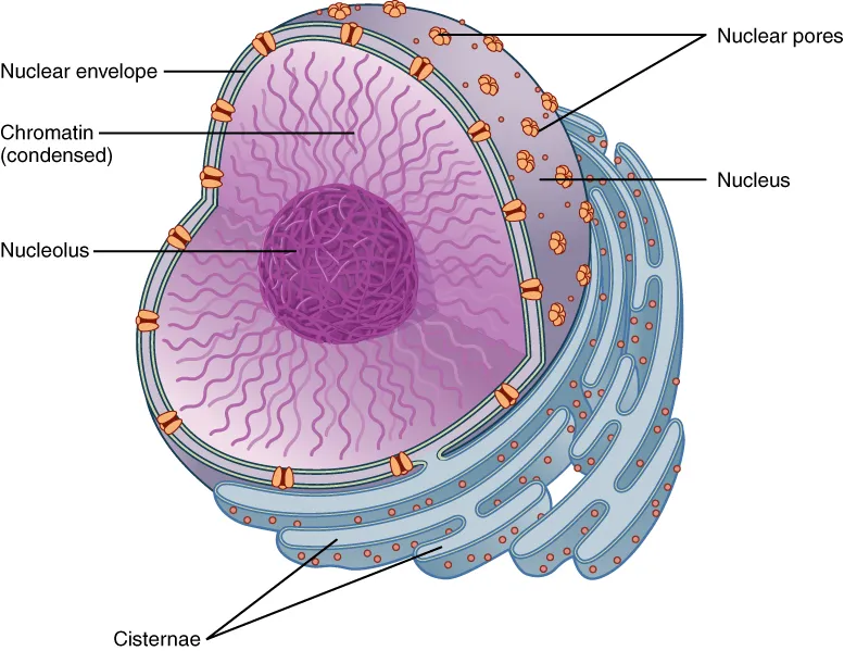
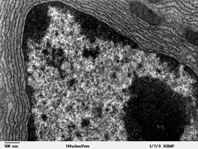
細胞核(直徑 5–10 µm)由雙層核膜包裹;外膜與粗麵內質網相連,也附有核糖體,兩層膜之間的間隙與內質網腔相通。內表面襯有由核纖層蛋白 A、B、C 構成的核纖層,既維持核的形狀,又錨定染色質。核纖層蛋白 A 突變導致一系列核纖層病,如埃默裡-德賴弗斯肌營養不良和哈欽森-吉爾福德早老症。[7][8]
細胞核與細胞質之間的所有物質交通都經過核孔:核孔複合體約 110 MDa,由約 30 種核孔蛋白、總計約 1,000 個複製按八重對稱組裝而成。通道內是富含苯丙胺酸-甘胺酸的蛋白鏈網,小分子自由擴散,而大分子蛋白和信使 RNA 必須由 Ran-GTP 梯度驅動的轉運受體(輸入蛋白、輸出蛋白)攜帶。一個細胞核有數千個核孔。[9][10][11]
約 2 m 長的 DNA 纏繞在核小體上,再經多級摺疊:染色質環、拓撲關聯結構域(TAD)、活性與非活性區室,最後每條染色體佔據各自的染色體疆域。緻密、轉錄沉默的異染色質傾向於貼附核纖層和核仁周圍,而開放、活躍的常染色質佔據核內部。核仁在核糖體 RNA 基因簇周圍無膜形成,在此轉錄 rRNA、組裝核糖體亞基,然後經核孔輸出。[12][13][14][15]
內質網、高基氏體與分泌途徑
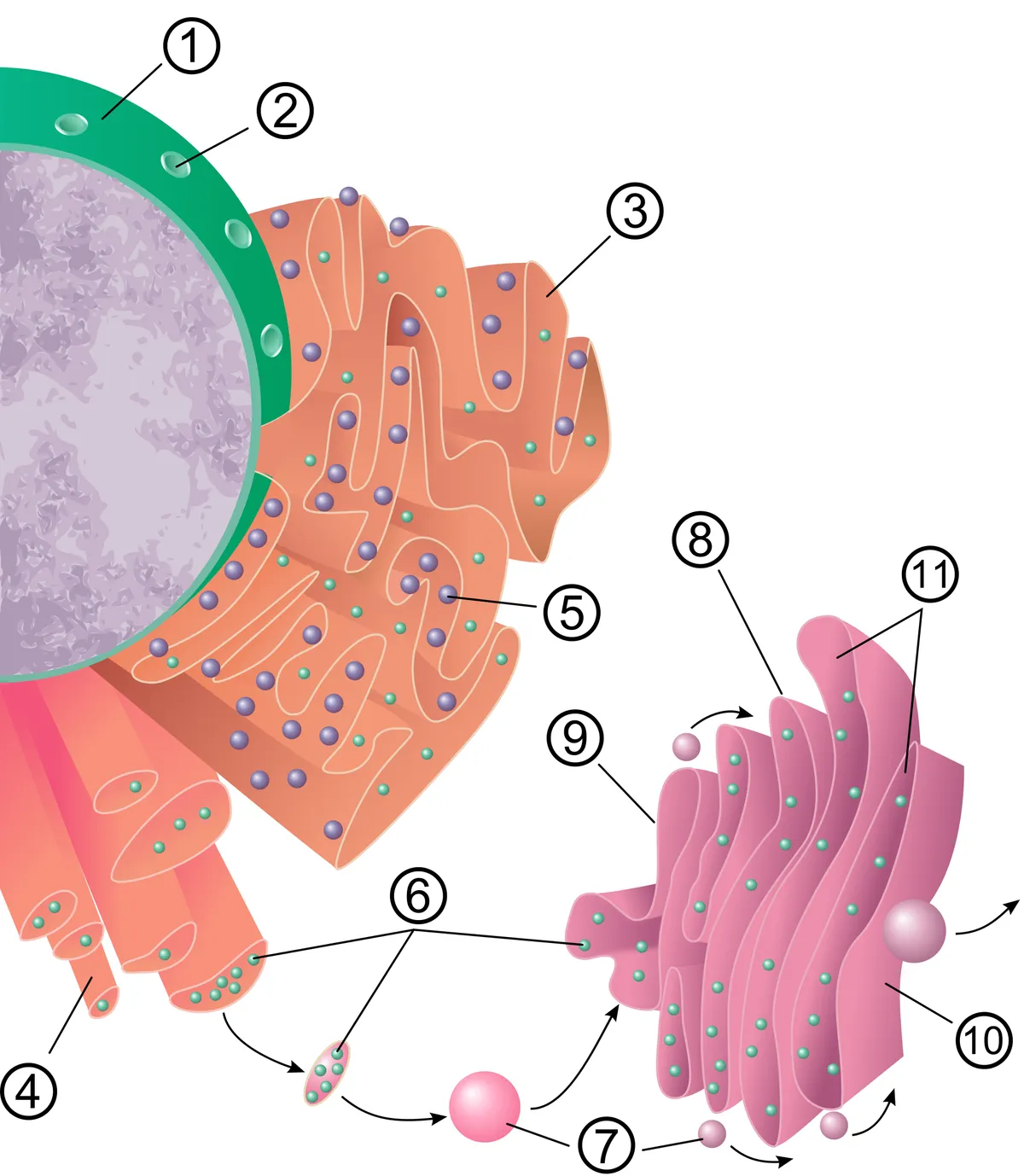
內質網是細胞內最大的連續膜系統,由與核膜相連的扁平片層和小管構成。粗麵內質網是外表面附有核糖體的層疊扁平池:分泌蛋白、膜蛋白和溶體酶直接被翻譯進腔內,在那裡由分子伴侶摺疊、形成二硫鍵並加上最初的 N-連線糖鏈。錯誤摺疊的蛋白被滯留並降解;當它們堆積時,細胞啟動未摺疊蛋白反應。[16][17]
滑面內質網是無核糖體的小管網路,合成磷脂和膽固醇、類固醇激素(腎上腺皮質、睪丸),以細胞色素 P450 酶解毒藥物(肝細胞),並儲存 Ca²⁺;在肌細胞中它成為肌質網。內質網還在間距 10–30 nm 的膜接觸位點與粒線體、質膜、內體和脂滴相接,不經囊泡直接傳遞脂質和 Ca²⁺。[18][19][20]
蛋白質以 COPII 包被囊泡離開內質網,前往高基氏體。高基氏體由 4–8 個扁平池極性排列而成:順面接收貨物,反面發出貨物。貨物通過時,從順面到反面依次排列的糖基轉移酶依序修剪和延長糖鏈,溶體酶則被加上甘露糖-6-磷酸標籤。在反面高基氏網,貨物被分入三條路線:送往溶體的囊泡、通向質膜的組成型分泌,以及儲存至訊號到來才釋放的調節型分泌顆粒(胰島素、胰酶)。[21][22][23][24]
粒線體
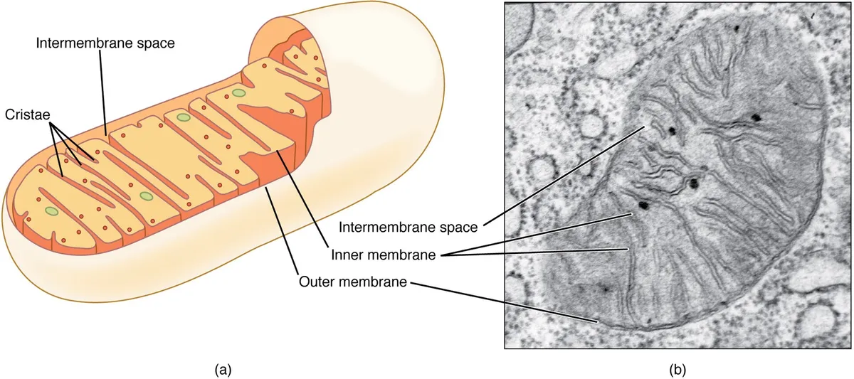
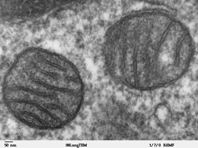
粒線體有兩層膜:對小分子通透的光滑外膜,和摺疊成嵴、含有電子傳遞鏈和 ATP 合酶的內膜。跨內膜的質子梯度驅動 ATP 合酶,提供細胞大部分 ATP;基質中含三羧酸迴圈和脂肪酸氧化的酶、環狀粒線體 DNA(37 個基因)及其自身的核糖體。粒線體數量與能量需求相稱:在心肌細胞中約佔三分之一體積。[25][26][27]
粒線體並非固定的單個顆粒,而是不斷融合與分裂的動態網路。融合(外膜的粒線體融合蛋白、內膜的 OPA1)使蛋白質和 DNA 共享;分裂由動力蛋白 DRP1 在與內質網的接觸位點縊縮完成,把受損部分分離出來,經粒線體自噬清除。約 99% 的粒線體蛋白由核基因編碼,經 TOM 和 TIM 複合體輸入。[28][29][30][31]
除供能外,粒線體還緩衝 Ca²⁺,產生用作訊號的活性氧,並決定程式性細胞死亡:外膜通透化後,細胞色素 c 逸出並激活半胱天冬酶。釋放到胞質中的粒線體 DNA 被識別為外來物而引發炎症。粒線體形態和動力學紊亂見於神經退行性疾病、心力衰竭和腎病。[32][33][34]
溶體、自噬與過氧化體
溶體是單層膜囊泡,其酸性腔(pH 約 4.5–5,由 V-ATP 酶質子泵維持)含 50 多種水解酶。物質通過三條途徑到達:內吞(質膜的受體和液體經早期、晚期內體)、吞噬(巨噬細胞中的細菌和死細胞)和自噬。從內體到溶體的成熟由 GTP 酶 Rab5、隨後 Rab7 調控。缺少某一種溶體酶會導致貯積病,如戈謝病或泰-薩克斯病。[35][36]
自噬時,雙層膜包裹一部分胞質、蛋白聚集體或受損細胞器形成自噬體,再與溶體融合,消化並回收產物。自噬在飢餓時至關重要,也是神經元的主要質量控制系統,因為神經元不能靠分裂稀釋廢物;自噬-溶體途徑失靈參與阿茲海默症、帕金森氏症和亨廷頓病。[37][38][39]
過氧化體是小型(0.2–1 µm)單層膜囊泡,含產生 H₂O₂ 的氧化酶和分解它的過氧化氫酶。它們對極長鏈脂肪酸進行 β 氧化、對植烷酸進行 α 氧化,並合成縮醛磷脂(髓鞘的關鍵脂質)和膽汁酸。其蛋白由過氧化體生物發生因子(PEX 基因)從胞質輸入,缺陷導致齊薇格綜合徵等過氧化體生物發生障礙。[40][41][42]
細胞骨架
細胞骨架由三種纖維系統組成。肌動蛋白微絲(7 nm)在膜下皮層最密集,構成微絨毛、片狀偽足和胞質分裂環,並與肌球蛋白共同產生力。微管(25 nm)是由 α/β 微管蛋白構成的中空管,從中心體以正端向外生長;它們不斷生長和塌縮(“動態不穩定性”),充當驅動蛋白(向外周)和動力蛋白(向中心)的軌道,並組成有絲分裂紡錘體。[43]
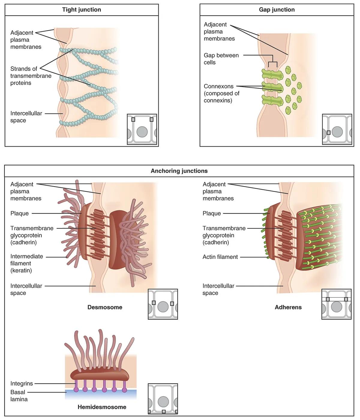
中間絲(10 nm)機械強度高且具有細胞型別特異性:上皮的角蛋白、間充質細胞的波形蛋白、肌肉的結蛋白、星狀膠細胞的 GFAP、神經元的神經絲以及核內的核纖層蛋白。它們附著於橋粒和半橋粒,把張力分散到整個組織;角蛋白突變使皮膚輕微摩擦即起水皰。由於具有細胞型別特異性,它們被用作腫瘤標誌物,血中神經絲是軸突損傷的生物標誌物。[44][45][46]
中心體由兩個相互垂直的中心粒組成,中心粒是直徑約 250 nm、長 500 nm 的圓筒,壁由九組三聯微管構成,周圍是成核微管的中心粒周圍物質。中心粒也作為纖毛的基體:纖毛軸絲為 9 + 2 結構,由動力蛋白驅動擺動。氣道的多纖毛細胞帶有數百根同步擺動的纖毛推動黏液;纖毛運動障礙導致反覆支氣管炎、鼻竇炎和不育。[47][48][49]
細胞週期與細胞分裂
細胞週期包括間期(G1、複製 DNA 的 S 期、G2)和 M 期。由細胞週期蛋白-CDK 複合體控制的檢查點只在 DNA 完整且完全複製時才讓細胞繼續前進;轉錄因子 MYC 推動細胞進入週期,在許多癌症中過度活躍。神經元等高度分化細胞永久退出週期進入 G0;衰老細胞永久停止分裂,但持續分泌炎症因子。[50][51]
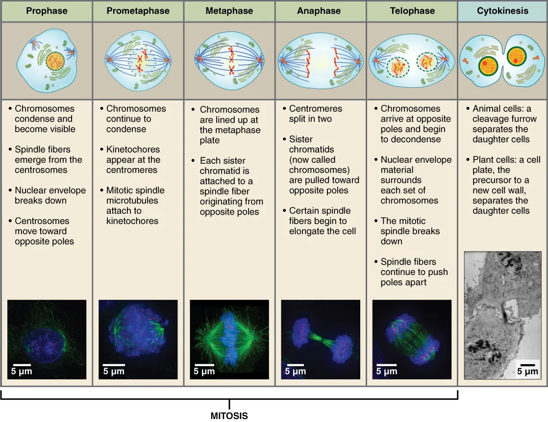
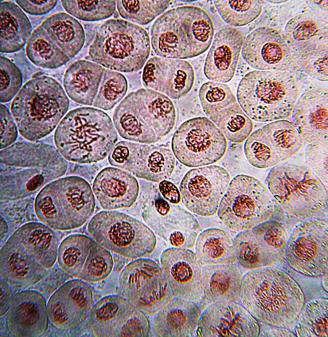
有絲分裂時,染色體凝縮,核膜解體(前期、前中期),紡錘體微管通過著絲粒上結合微管末端的蛋白複合體——動粒——捕獲著絲粒。紡錘體檢查點使細胞停在中期,直到每條染色體都與兩極連線;隨後黏連蛋白被切開,姐妹染色單體分向兩極(後期),核膜重建(末期),肌動球蛋白環把胞質一分為二。分離錯誤產生非整倍體,這是流產的主要原因,也是許多腫瘤的特徵。[52][53][54][55]
全文閱讀(StatPearls)
本主題的 StatPearls 章節按原文收錄。其 CC BY-NC-ND 4.0 授權僅允許不加修改的複製,因此不作翻譯或刪節。
Anatomy, Back, Nucleus PulposusNedresky D, Reddy V, Singh G · StatPearls 2023 · NBK535373 · 8k 字元
正在載入…
Nedresky D, Reddy V, Singh G. Anatomy, Back, Nucleus Pulposus. StatPearls [Internet]. Treasure Island (FL): StatPearls Publishing; 2023. NBK535373 · PMID 30570994 · CC BY-NC-ND 4.0. 按原文收錄,未作修改。
Histology, Rough Endoplasmic ReticulumSanvictores T, Davis DD · StatPearls 2023 · NBK563126 · 7k 字元
正在載入…
Sanvictores T, Davis DD. Histology, Rough Endoplasmic Reticulum. StatPearls [Internet]. Treasure Island (FL): StatPearls Publishing; 2023. NBK563126 · PMID 33085273 · CC BY-NC-ND 4.0. 按原文收錄,未作修改。
本系統的器官
參考文獻
本頁數值與描述的來源,收錄於 PubMed(美國國家醫學圖書館)。
- [1]Essay on Biomembrane StructureGerle C, The Journal of membrane biology 2019PMID 30877332
- [2]Lipid Rafts: Controversies Resolved, Mysteries RemainLevental I et al., Trends in cell biology 2020PMID 32302547
- [3]Lipid rafts as a membrane-organizing principleLingwood D et al., Science 2010PMID 20044567
- [4]CaveolaeParton RG et al., Current biology 2018PMID 29689223
- [5]The endothelial glycocalyx: composition, functions, and visualizationReitsma S et al., Pflugers Archiv 2007PMID 17256154
- [6]The glycocalyx: a novel diagnostic and therapeutic target in sepsisUchimido R et al., Critical care 2019PMID 30654825
- [7]Lamina-associated domains: Tethers and loosenersManzo SG et al., Current opinion in cell biology 2022PMID 35189475
- [8]Shared mechanisms in physiological and pathological nucleoplasmic reticulum formationDrozdz MM et al., Nucleus 2017PMID 27797635
- [9]The Structure of the Nuclear Pore Complex (An Update)Lin DH et al., Annual review of biochemistry 2019PMID 30883195
- [10]Structure, function and assembly of nuclear pore complexesPetrovic S et al., Nature reviews. Molecular cell biology 2026PMID 40926106
- [11]Aggregation, Phase Separation and Spatial Morphologies of the Assemblies of FG NucleoporinsZilman A, Journal of molecular biology 2018PMID 30017917
- [12]Chromosome territories and the global regulation of the genomeFritz AJ et al., Genes, chromosomes & cancer 2019PMID 30664301
- [13]Decoding the organization, dynamics, and function of the 4D genomeAboelnour E et al., Developmental cell 2021PMID 33984271
- [14]The nucleolusPederson T, Cold Spring Harbor perspectives in biology 2011PMID 21106648
- [15]Eukaryotic Ribosome AssemblyVanden Broeck A et al., Annual review of biochemistry 2024PMID 38768392
- [16]Histology, Rough Endoplasmic ReticulumSanvictores T et al., StatPearls 2023PMID 33085273
- [17]The endoplasmic reticulum: structure, function and response to cellular signalingSchwarz DS et al., Cellular and molecular life sciences 2016PMID 26433683
- [18]Structure and function of ER membrane contact sites with other organellesPhillips MJ et al., Nature reviews. Molecular cell biology 2016PMID 26627931
- [19]Endoplasmic reticulum-mitochondria contacts: function of the junctionRowland AA et al., Nature reviews. Molecular cell biology 2012PMID 22992592
- [20]Zooming into lipid droplet biology through the lens of electron microscopyDudka W et al., FEBS letters 2024PMID 38726814
- [21]Making COPII coatsKirchhausen T, Cell 2007PMID 17604713
- [22]The Golgi complex: a hub of the secretory pathwayPark K et al., BMB reports 2021PMID 33612152
- [23]Golgi glycosylationStanley P, Cold Spring Harbor perspectives in biology 2011PMID 21441588
- [24]Mechanistic insights into cargo sorting and export from the Golgi apparatusWatson ET et al., Nature reviews. Molecular cell biology 2025PMID 41214333
- [25]Mitochondrial proteins: from biogenesis to functional networksPfanner N et al., Nature reviews. Molecular cell biology 2019PMID 30626975
- [26]Structure and Function of the Mitochondrial RibosomeGreber BJ et al., Annual review of biochemistry 2016PMID 27023846
- [27]Kinetic Mathematical Modeling of Oxidative Phosphorylation in Cardiomyocyte MitochondriaTseng WW et al., Cells 2022PMID 36552784
- [28]Molecular mechanisms of mitochondrial dynamicsTábara LC et al., Nature reviews. Molecular cell biology 2025PMID 39420231
- [29]The cell biology of mitochondrial membrane dynamicsGiacomello M et al., Nature reviews. Molecular cell biology 2020PMID 32071438
- [30]Mitochondrial Machineries for Protein Import and AssemblyWiedemann N et al., Annual review of biochemistry 2017PMID 28301740
- [31]Mechanisms of mitophagyYoule RJ et al., Nature reviews. Molecular cell biology 2011PMID 21179058
- [32]Mitochondria and cell death-associated inflammationVringer E et al., Cell death and differentiation 2023PMID 36447047
- [33]Mitochondria in disease: changes in shapes and dynamicsJenkins BC et al., Trends in biochemical sciences 2024PMID 38402097
- [34]Mitochondrial dysfunction and the AKI-to-CKD transitionJiang M et al., American journal of physiology. Renal physiology 2020PMID 33073587
- [35]Rab GTPase Function in Endosome and Lysosome BiogenesisLangemeyer L et al., Trends in cell biology 2018PMID 30025982
- [36]Lysosomal quality control ReviewHenn D et al., Autophagy 2025PMID 39968899
- [37]Autophagy pathway: Cellular and molecular mechanismsYu L et al., Autophagy 2018PMID 28933638
- [38]Mechanisms of autophagy-lysosome dysfunction in neurodegenerative diseasesNixon RA et al., Nature reviews. Molecular cell biology 2024PMID 39107446
- [39]Autophagy genes in biology and diseaseYamamoto H et al., Nature reviews. Genetics 2023PMID 36635405
- [40]Peroxisome: Metabolic Functions and BiogenesisOkumoto K et al., Advances in experimental medicine and biology 2020PMID 33417203
- [41]Peroxisomes: a nexus for lipid metabolism and cellular signalingLodhi IJ et al., Cell metabolism 2014PMID 24508507
- [42]Alpha-oxidationJansen GA et al., Biochimica et biophysica acta 2006PMID 16934890
- [43]Microtubules and Microtubule-Associated ProteinsGoodson HV et al., Cold Spring Harbor perspectives in biology 2018PMID 29858272
- [44]Plasticity of cytoplasmic intermediate filament architecture determines cellular functionsSchwarz N et al., Current opinion in cell biology 2023PMID 37918274
- [45]GFAP at 50Messing A et al., ASN neuro 2020PMID 32811163
- [46]Neurofilaments as biomarkers in neurological disorders - towards clinical applicationKhalil M et al., Nature reviews. Neurology 2024PMID 38609644
- [47]Centriole structureWiney M et al., Philosophical transactions of the Royal Society of London. Series B, Biological sciences 2014PMID 25047611
- [48]Multiciliated cellsBrooks ER et al., Current biology 2014PMID 25291643
- [49]Towards an atomic model of a beating ciliary axonemeGrossman-Haham I, Current opinion in structural biology 2023PMID 36586349
- [50]Myc and cell cycle controlBretones G et al., Biochimica et biophysica acta 2015PMID 24704206
- [51]A guide to assessing cellular senescence in vitro and in vivoGonzález-Gualda E et al., The FEBS journal 2021PMID 32961620
- [52]PrometaphaseFerreira LT et al., Seminars in cell & developmental biology 2021PMID 34127384
- [53]The kinetochoreCheeseman IM, Cold Spring Harbor perspectives in biology 2014PMID 24984773
- [54]Mitotic genome foldingHirano T, The Journal of cell biology 2025PMID 40492990
- [55]Human aneuploidy: mechanisms and new insights into an age-old problemNagaoka SI et al., Nature reviews. Genetics 2012PMID 22705668